-
Modified Barium Swallow Study (MBSS) One Gold Standard Assessment
View : Modified Barium Swallow Study (MBSS) One Gold Standard AssessmentThe dysphagia field is divided between Modified Barium Swallow Study (MBSS) and Flexible Endoscopic Evaluation of Swallowing (FEES). Each assessment has its strengths, and using both together offers a comprehensive understanding of swallowing function. Advocating for access to both methods can enhance patient evaluation and treatment outcomes, focusing on physiological deficits rather than just safety.
-

30 Facts About Dysphagia to Raise Awareness and Improve Patient Care
View : 30 Facts About Dysphagia to Raise Awareness and Improve Patient CareJune is Dysphagia Awareness Month, highlighting the condition affecting millions. Dysphagia, or difficulty swallowing, can stem from various underlying issues and leads to serious health risks. Speech-Language Pathologists play a vital role in diagnosis and treatment. Raising awareness about dysphagia improves outcomes for patients, caregivers, and healthcare providers alike.
-
Nutrition Essentials for Modified Diets
View : Nutrition Essentials for Modified DietsNutrition is vital for health, especially for individuals on modified diets, who are at risk of malnutrition. Macronutrients (carbohydrates, proteins, fats) and micronutrients (vitamins, minerals) play crucial roles. Strategies such as food fortification, small frequent meals, and hydration can enhance nutritional intake, ensuring patients maintain health and well-being despite dietary restrictions.
-
Modern Dysphagia Cooking: Turn Family Favorites into Dysphagia-Friendly Dishes
View : Modern Dysphagia Cooking: Turn Family Favorites into Dysphagia-Friendly DishesSimply Thick has released a cookbook featuring dysphagia-friendly recipes compliant with IDDSI levels 4, 5, and 6. It includes simple pureeing techniques for common foods, alongside informative chapters on dysphagia and nutrition. This resource aids families, patients, and caregivers in preparing compliant meals, enhancing dietary understanding and meal preparation.
-

SWIK Oral Suction System
View : SWIK Oral Suction SystemThe SWIK Oral Suction System is an innovative solution for managing oral secretions. Unlike traditional suction methods, it uses a small sponge to collect secretions hands-free. Its design emphasizes user comfort and dignity. Created by hairdresser Rebecca Altounian, it offers a discreet alternative for those needing assistance.
-
Intensive Dysphagia Rehabilitation (IDR)
View : Intensive Dysphagia Rehabilitation (IDR)Intensive Dysphagia Rehabilitation (IDR) streamlines therapy for patients with moderate to severe dysphagia through evidence-based protocols leveraging neuroplasticity. By focusing on targeted swallowing exercises and patient adherence, IDR enhances treatment efficacy, improving patient outcomes and compliance. It marks a significant shift from compensation to active rehabilitation in dysphagia therapy.
-
Head and Neck Cancer and Dysphagia: Treatment Options and Timing
View : Head and Neck Cancer and Dysphagia: Treatment Options and TimingEffective management of dysphagia in head and neck cancer patients involves early assessment and initiation of swallowing therapy alongside treatment. Various strategies, including rigorous swallowing exercises and innovative programs like MD Anderson’s Swallowing Boot Camp, show promise in improving swallowing function and quality of life, despite challenges in standardization and adherence.
-

Math and Dysphagia
View : Math and DysphagiaThe blog post by George Barnes and Doreen Benson emphasizes the importance of incorporating statistics into speech-language pathology (SLP) practices, specifically for treating dysphagia. It discusses common cognitive biases affecting clinical decision-making and stresses the need for a more analytical approach in evaluating patient risks and benefits, promoting better outcomes in care.
-
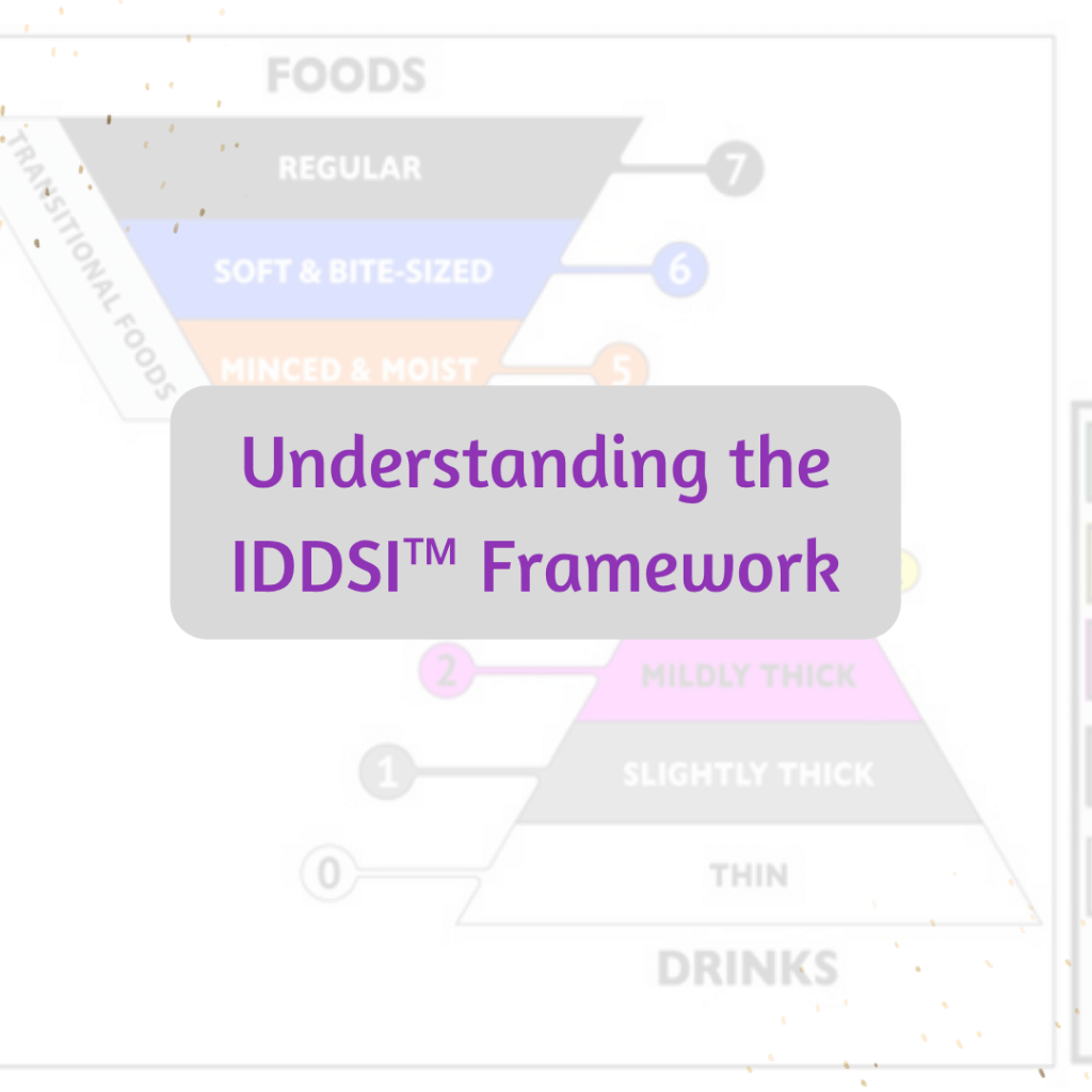
Understanding the IDDSI™ Framework
View : Understanding the IDDSI™ FrameworkThe International Dysphagia Diet Standardisation Initiative (IDDSI™) provides a unified system for food and drink consistencies, enhancing patient safety. It replaces the National Dysphagia Diet, offering clear levels for drinks and foods, minimizing risks. Resources such as testing cards and guides support implementation and training in diverse healthcare settings.
-

Measuring Outcomes for Success…..What are You Using?
View : Measuring Outcomes for Success…..What are You Using?The Dysphagia Toolbox offers valuable tools for dysphagia assessment and treatment. Various free outcome measures like questionnaires and screening tools support clinicians in evaluating patients effectively. These assessments track progress and ensure reliable evaluations, which are essential for optimal dysphagia management and documentation in compliance with healthcare standards.
The Clinician’s Place for Information, Ideas and Awareness


Popular posts

About the Author
Tiffani Wallace, author and creator behind Dysphagia Ramblings, is passionate about sharing information about dysphagia.